Fil:Nematocyst discharge.png
Det finns ingen version med högre upplösning.
Nematocyst_discharge.png (480 × 371 pixlar, filstorlek: 190 kbyte, MIME-typ: image/png)
| Denna fil tillhandahålls av Wikimedia Commons. Informationen nedan är kopierad från dess filbeskrivningssida. |

|
Denna bild (eller alla bilder i denna artikel eller kategori) bör återskapas med hjälp av vektorgrafik som en SVG-fil. Detta har flera fördelar; se Commons:Media for cleanup för mer information. Om det redan existerar en SVG-version av denna bild, var vänlig och lägg upp den. Efter att en SVG-version lagts upp, ersätt denna mall med {{vector version available|nytt bildnamn.svg}} på denna sida.
|
Sammanfattning
| Beskrivning |
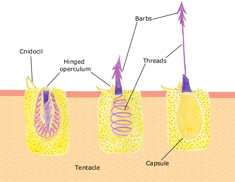
English: The diagram above shows the anatomy of a nematocyst cell and its “firing” sequence, from left to right. On the far left is a nematocyst inside its cellular capsule. The cell’s thread is coiled under pressure and wrapped around a stinging barb. When potential prey makes contact with the tentacles of a polyp, the nematocyst cell is stimulated. This causes a flap of tissue covering the nematocyst—the operculum—to fly open. The middle image shows the open operculum, the rapidly uncoiling thread and the emerging barb. On the far right is the fully extended cell. The barbs at the end of the nematocyst are designed to stick into the polyp’s victim and inject a poisonous liquid. When subdued, the polyp’s tentacles move the prey toward its mouth and the nematocysts recoil back into their capsules. |
| Datum | 11 april 2007 (ursprungligt uppladdningsdatum) |
| Källa | Överförd från en.wikipedia till Commons. |
| Skapare | The original uploader was Spaully på engelska Wikipedia. |
Licensiering
Den här filen är licensierad under Creative Commons Dela Lika 1.0.
Creative Commons har pensionerat det här juridiska verktyget och rekommenderar inte att det används för verk.
|
This image is in the public domain because it contains materials that originally came from the U.S. National Oceanic and Atmospheric Administration, taken or made as part of an employee's official duties.
|
Ursprunglig uppladdningslogg
Den ursprungliga beskrivningssidan fanns här. Alla följande användarnamn finns på en.wikipedia.
- 2007-04-11 17:10 Spaully 480×371×8 (194868 bytes) Modified from: http://www.oceanservice.noaa.gov/education/kits/corals/media/supp_coral01b.html {{Information |Description=Nematocyst discharge process. |Source= Modified from [http://www.oceanservice.noaa.gov/education/kits/corals/media/supp_coral01b.html
Filhistorik
Klicka på ett datum/klockslag för att se filen som den såg ut då.
| Datum/Tid | Miniatyrbild | Dimensioner | Användare | Kommentar | |
|---|---|---|---|---|---|
| nuvarande | 13 oktober 2007 kl. 19.29 | | 480 × 371 (190 kbyte) | Alison | {{Information |Description===Description== The diagram above shows the anatomy of a nematocyst cell and its “firing” sequence, from left to right. On the far left is a nematocyst inside its cellular capsule. The cell’s thread is coiled under pressur |
Filanvändning
Följande sida använder den här filen:
Global filanvändning
Följande andra wikier använder denna fil:
- Användande på ca.wikipedia.org
- Användande på ceb.wikipedia.org
- Användande på en.wikipedia.org
- Användande på fr.wikipedia.org
- Användande på hr.wikipedia.org
- Användande på id.wikipedia.org
- Användande på it.wikibooks.org
- Användande på ja.wikipedia.org
- Användande på lv.wikipedia.org
- Användande på ms.wikipedia.org
- Användande på my.wikipedia.org
- Användande på pa.wikipedia.org
- Användande på pt.wikipedia.org
- Användande på simple.wikipedia.org
- Användande på te.wikipedia.org
- Användande på th.wikipedia.org
- Användande på vi.wikipedia.org